


LavaCell™
LavaCell™ is a small, neutral, non-toxic compound that readily diffuses into live or fixed cells and spontaneously reacts to produce bright orange fluorescence. This naturally fluorescent compound is excited by a variety of common lasers and can be multiplexed with a wide range of common blue, green and yellow-emitting probes.
Because LavaCell is a neutral molecule that crosses cell membranes, cells require no pre-treatment or permeabilization prior to staining. LavaCell rapidly reacts with free amine groups on proteins, which produces a bright orange fluorescent signal (Figure 1). LavaCell does not stain nucleic acids nor does it adversely affect cell growth. LavaCell can be excited with common laser sources (405, 488 and 532 nm) and has a Stokes shift of over 100 nm with peak fluorescence emission at 610 nm.
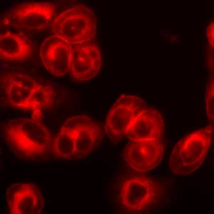
Figure 1: Staining of live CHO cells with LavaCell.
LavaCell stains the plasma membrane and internal membranes, such as those of the nucleus, Golgi and ER. CHO cells were plated in 96-well plates cells and treated with 6 µM LavaCell (diluted in complete medium) for 30 minutes at 37°C. After loading, the dye containing DMEM was removed and cells were washed once in PBS. Cells were imaged in PBS using a fluorescent microscope with a 540-580 nm excitation filter and a 600-660 nm emission filter (exposure time 10 seconds, 40X objective).
LavaCell advantages
- Water soluble fluorescent cell stain
- Fluoresces on interaction with proteins; no need to wash away unbound stain
- Readily permeates into cells
- No effect on cell growth or viability
- Ideal for multiplexing with other fluorophores due to long Stokes shift
LavaCell™ is a small dye that readily diffuses into live or fixed cells, where it stains the plasma membrane and internal membranes such as the Endoplasmic Reticulum, the Golgi and the nuclear envelope. This staining pattern has been shown in a number of different cell-lines (Figures 2 and 3).
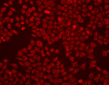
Figure 2: Staining of live HeLa cells with LavaCell.
HeLa cells were plated in 96-well plates cells and treated with 6 µM LavaCell (diluted in complete medium) for 30 minutes at 37°C. After loading, the media/LavaCell was removed and the cells were washed once in PBS. The cells were then imaged in PBS using a fluorescent microscope with a 540-580 nm excitation filter and a 600-660 nm emission filter.
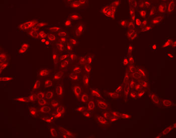
Figure 3: Staining of live U2OS cells with LavaCell.
U2OS osteosarcoma cells were plated in 96-well plates cells and treated with 6 µM LavaCell (diluted in complete medium) for 30 minutes at 37°C. After loading, the media/LavaCell was removed and the cells were washed once in PBS. The cells were then imaged in PBS using a fluorescent microscope with a 540-580 nm excitation filter and a 600-660 nm emission filter.
LavaCell, which can be excited with commonly used light sources between 405 and 532 nm, has its emission peak at 610 nm. This broad Stokes shift not only effectively separates the fluorescent emission from the excitation wavelength, it enables LavaCell to be used in co-staining with other cellular stains or other fluorescent dyes (Figure 4).
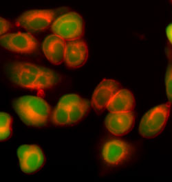
Figure 4: Co-staining of live CHO cells with LavaCell and calcein AM.
CHO cells were plated in 96-well plates and treated with LavaCell and the live cell stain calcein AM (diluted in complete medium) for 30 minutes at 37°C. After loading, the media/LavaCell was removed and the cells were washed once in PBS. The cells were then imaged in PBS using a fluorescent microscope with the following filter sets: a 540-580 nm excitation filter and a 600-660 nm emission filter for LavaCell and a combination of a 488 nm excitation filter and a 500-550 nm emission filter for calcein AM. The LavaCell stains the internal and external membranes an orange/red color, while calcein AM is the green color seen throughout the cell, including the nucleus and cytosol.
In addition to fluorescent microscopy, LavaCell has been shown to work in fluorescent plate-based assays for monitoring cell coverage of cell culture plates or individual wells of a 96-well plate. In addition, LavaCell can be used in High-throughput Screening (HTS), where it can be used to monitor changes in cell proliferation of the entire cell population (Figure 5), or proliferation of individual clones in cell colony growth assays.

