
PARP and DDR Pathway Drug Discovery
DNA damage response (DDR) is a normal cellular function that can result in DNA repair, cell cycle arrest, senescence and/or apoptosis in response to cellular DNA damage.

Defects in the DDR pathway lead to genomic instability, cancer initiation and uncontrolled cell proliferation. Many kinases such as WEE1, DNA PK and ATM/ATR in the DDR pathway have been pursued as drug targets for cancer therapy. The 17 member Poly (ADP ribose) Polymerase (PARP) family of proteins mediate a number of cellular processes, including gene transcription and DNA repair, and are widely implicated in human disease.
Interested in more information about PARP/DDR pathway drug discovery solutions? Contact us! info@mybio.ie
Numerous signaling networks are associated with DDR mechanisms. These modes involve sensors (e.g., PARPs), mediators (e.g., DNA PK, ATM, ATR), and transducers and effectors of signaling (e.g., kinases). The PARP family of proteins is classified in two groups based on poly vs. mono ADP ribosylation.


PARP Target Engagement
NanoBRET™ technology offers a sensitive, specific method to measure the interaction of small-molecule drugs with their target protein in live cells.
With the NanoBRET™ Target Engagement Assay, you can:
- Quantitate compound affinity (how tightly it binds to a protein) and target protein occupancy (how much compound binds to a protein) in live cells.
- Assess how long a compound binds to the target protein (its residence time) under physiological conditions.
- Scale the simple, multiwell assay to suit your research throughput needs.
- Generate high‐quality data with low error rates and high reproducibility.
- Get started quickly with available ready-to-use assays.

Principle of the NanoBRET™ Target Engagement Assay. Binding of a test compound results in loss of NanoBRET™ signal between a target-NanoLuc® fusion protein and a cell-permeable fluorescent NanoBRET™ Tracer inside intact cells.
Measure PARP Target Engagement in Live Cells
PARP1 inhibitors are promiscuous against lesser studied PARPs.
FDA-approved and clinical PARP Inhibitors engage a spectrum of mono-PARPs in cells.


Measure PARP Activity Using the NAD/NADH-Glo™ Assay
Upon DNA damage, PARP activity is upregulated. Monomeric NAD(+) is consumed and enzymatically polymerized into a Poly ADP ribosylation chain at the site of the damage or a mono ADP ribose added to the target substrate.
The bioluminescent NAD/NADH-Glo™ Assay is a homogeneous assay that detects NAD(+) and NADH and determines their ratios in biological samples such as cell lysates. The simple add-and-read format can be easily adapted for high-throughput screen. A modified protocol is available upon request for measuring activity of PARPs and other enzymes.

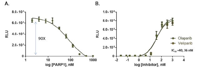
PARP activity assay. PARP1 activity was measured in reactions containing PARP1, 375ng nucleosomes and 20μM NAD in a 384-well plate. After a 1-hour incubation, NAD levels were analyzed by adding an equal volume of modified NAD/NADH Glo™ Detection Reagent. Luminescence was recorded after 20 minutes. Panel A: Titration of PARP1 enzyme (in 5μl volume). Panel B: PARP1 inhibition.
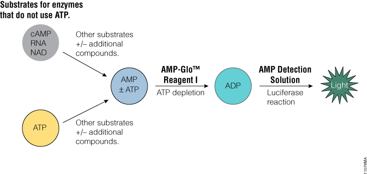
AMP-Glo™ Assay to Measure PARP Activity
The AMP-Glo™ Assay is a homogeneous assay that generates a luminescent signal from any biochemical reaction that produces AMP. It can be used to measure the activity of a broad range of enzymes, such as cyclic AMP specific phosphodiesterases , aminoacyl tRNA synthetases, DNA ligases and ubiquitin ligases or enzymes modulated by AMP.
Read a publication using this assay to monitor Poly ADP ribosylation: Mondal, S., Hsiao, K. and Goueli, S.A. (2017) Utility of Adenosine Monophosphate Detection System for Monitoring the Activities of Diverse Enzyme Reactions. ASSAY and Drug Development Technologies 15, 300–341.
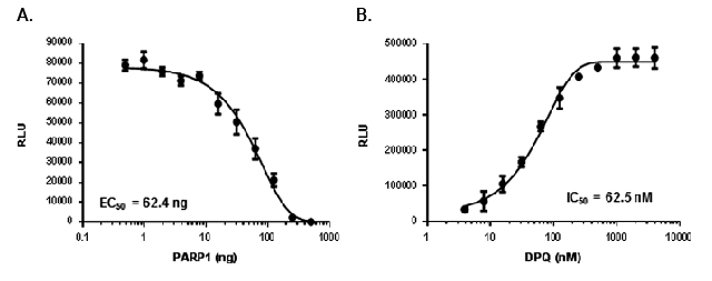
Measurement of ADPribosylation using the modified AMP-Glo™ assay. Panel A: Poly ADP ribosylation of PARP1 on nucleosomes. Reactions containing PARP1 activity was measured using 50μg/ml nucleosomes and 20μM NAD ++, and different amounts of PARP1 were analyzed by the AMP-Glo™ system. Panel B: Detection of PARP1 inhibition by DPQ.
Kinases in DDR
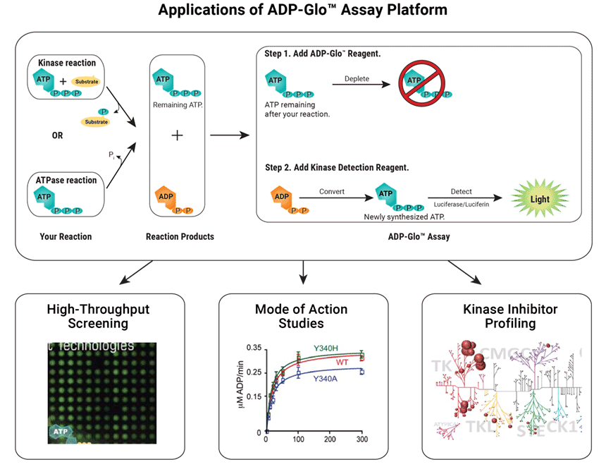
Kinase Selectivity Profiling with the ADP-Glo™ Assay
The ADP-Glo™ Kinase Assay is a Universal in vitro Biochemical Assay for all types of kinase studies. The assay measures ADP formed from a kinase reaction. The ADP is converted into ATP, which is used to generate a stable luminescent signal.
Applications of the ADP-Glo™ Assay include:
- High-throughput screening
- Mode of action studies
- Kinase inhibitor profiling
Kinase Enzyme Systems, which are optimized for use with the ADP-Glo™ Assay, allow you to easily screen and profile kinase inhibitors. The following kinases related to the DNA Damage Response are available:
- DNA-PK
- CHK1
- CHK2
- CDK2/Cyclin A1
- CDK2/Cyclin E1
- CDK6/Cyclin D3
- PLK-family
- CK2α isoforms
- MAPKAPK2 (MK2)
- MARK3
Live-Cell Target Engagement
The NanoBRET™ Target Engagement (TE) Kinase Assays allow you to quantitate compound binding to full length kinases inside live cells. Assays are available for >340 kinases with individual application notes for each kinase, including many kinases involved in DDR (e.g., CHK1, CHK2, CDK2/ Cyclin A1, CDK2/Cyclin E1, CDK6/Cyclin D, PKMYT1, PLK family, CK2 α isoforms, MAPKAPK2 (MK2), MARK3, Wee1). Individual application notes are available for each kinase. View the Kinase Target Engagement Assay Selection Guide for a complete list of available kinases.


DDR kinase inhibitor selectivity in live cells, compared to cell-free analysis.
Detection of Total or Phosphorylated Cellular Targets
Study Signaling Nodes in DNA Damage Response Using Lumit™ Immunoassay Cellular Systems
The Lumit™ Immunoassay Cellular System is a no-wash bioluminescent immunoassay that measures target analytes directly in cell lysates. The assay can be customized to detect nodes of signaling involved in DNA damage response using a simple workflow that doesn’t require media remove or lysate transfer. Simply add labeled antibodies to the sample, add the detection reagent and read the luminescent signal—all in a single plate.


